Medical Disclaimer: This is educational content only, not medical advice. Consult a licensed healthcare provider for diagnosis/treatment. Information based on sources like WHO/CDC guidelines (last reviewed: 2026-02-13).
This article is being expanded for more depth. Check back soon!
Pharyngeal Arches Clefts and Pouches Explained With Derivatives and Clinical Correlation
Frequently Asked Questions
What are pharyngeal arches?
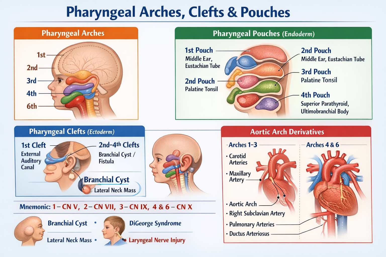
Pharyngeal arches are paired embryological structures that appear during the fourth week of development and give rise to the bones, muscles, nerves, and arteries of the face, neck, pharynx, and larynx.
How many pharyngeal arches are present in humans?
Six pairs of pharyngeal arches develop, but the fifth arch is rudimentary and disappears, so only arches 1, 2, 3, 4, and 6 contribute to adult structures.
What components does each pharyngeal arch contain?
Each pharyngeal arch contains a cartilage component, a muscular component, a cranial nerve, and an arterial (aortic arch) component.
Which nerve is associated with the first pharyngeal arch?
The first pharyngeal arch is supplied by the trigeminal nerve, mainly its mandibular division.
Which pharyngeal arch forms muscles of facial expression?
The second pharyngeal arch forms the muscles of facial expression and is supplied by the facial nerve.
Which muscle is the main derivative of the third pharyngeal arch?
The stylopharyngeus muscle is the primary derivative of the third pharyngeal arch.
Which cranial nerve supplies the third pharyngeal arch?
The third pharyngeal arch is supplied by the glossopharyngeal nerve.
Which arches are supplied by the vagus nerve?
The fourth and sixth pharyngeal arches are supplied by the vagus nerve via its laryngeal branches.
What are pharyngeal clefts?
Pharyngeal clefts are ectoderm-lined grooves located externally between adjacent pharyngeal arches.
Which pharyngeal cleft persists in adults?
Only the first pharyngeal cleft persists and forms the external auditory canal and part of the tympanic membrane.
What is a branchial cleft cyst?
A branchial cleft cyst is a congenital lateral neck swelling caused by persistence of the second pharyngeal cleft.
What are pharyngeal pouches?
Pharyngeal pouches are endoderm-lined outpouchings from the primitive pharynx that form internal structures of the head and neck.
What does the first pharyngeal pouch form?
The first pharyngeal pouch forms the middle ear cavity and the auditory (Eustachian) tube.
Which pharyngeal pouch forms the palatine tonsil?
The second pharyngeal pouch forms the palatine tonsil and tonsillar fossa.
Which pharyngeal pouch gives rise to the thymus?
The ventral wing of the third pharyngeal pouch gives rise to the thymus.
Which pharyngeal pouches form the parathyroid glands?
The third pouch forms the inferior parathyroid glands, while the fourth pouch forms the superior parathyroid glands.
What is the ultimobranchial body?
The ultimobranchial body is derived from the fourth pharyngeal pouch and forms the parafollicular C cells of the thyroid gland.
What is DiGeorge syndrome related to pharyngeal pouches?
DiGeorge syndrome results from failure of development of the third and fourth pharyngeal pouches, leading to thymic aplasia and hypocalcemia.
Which pharyngeal arch forms intrinsic laryngeal muscles?
Intrinsic muscles of the larynx are derived mainly from the sixth pharyngeal arch.
Why are pharyngeal arches clinically important?
Abnormal development of pharyngeal arches, clefts, or pouches leads to congenital anomalies such as branchial cysts, facial deformities, ear defects, and immune disorders.
MCQ Test - Pharyngeal Arches Clefts and Pouches Explained With Derivatives and Clinical Correlation
Progress:
0/0
Time: 00:00
No MCQs available for this article.