Medical Disclaimer: This is educational content only, not medical advice. Consult a licensed healthcare provider for diagnosis/treatment. Information based on sources like WHO/CDC guidelines (last reviewed: 2026-02-13).
This article is being expanded for more depth. Check back soon!
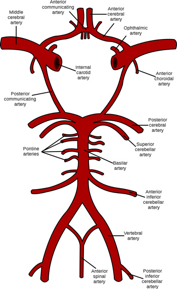
Blood Supply of the Brain Anatomy Arteries Circle of Willis and Clinical Correlation
Frequently Asked Questions
What is the main blood supply of the brain?
The brain is supplied by two main arterial systems: the internal carotid arteries forming the anterior circulation and the vertebrobasilar system forming the posterior circulation.
Which artery is most commonly involved in ischemic stroke?
The middle cerebral artery is the most commonly involved artery in ischemic stroke due to its large size and direct continuation from the internal carotid artery.
What is the Circle of Willis?
The Circle of Willis is an arterial anastomotic ring at the base of the brain that connects the internal carotid and vertebrobasilar systems and provides collateral circulation.
Which areas of the brain are supplied by the anterior cerebral artery?
The anterior cerebral artery supplies the medial surfaces of the frontal and parietal lobes including the motor and sensory areas for the contralateral lower limb.
Which artery supplies the visual cortex?
The posterior cerebral artery supplies the occipital lobe and primary visual cortex.
What are lenticulostriate arteries and why are they clinically important?
Lenticulostriate arteries are small penetrating branches of the middle cerebral artery that supply the basal ganglia and internal capsule and are prone to hypertensive hemorrhage and lacunar infarcts.
Which arteries supply the brainstem and cerebellum?
The vertebrobasilar system including the vertebral arteries and their branches such as PICA AICA SCA and basilar artery supplies the brainstem and cerebellum.
What is cerebral autoregulation?
Cerebral autoregulation is the ability of cerebral blood vessels to maintain constant blood flow despite changes in systemic blood pressure typically between mean arterial pressures of 60 to 160 mmHg.
What causes berry aneurysms and where do they occur?
Berry aneurysms are caused by congenital weakness in arterial walls and most commonly occur at bifurcation points of arteries in the Circle of Willis.
How does venous blood drain from the brain?
Venous blood from the brain drains through superficial and deep cerebral veins into dural venous sinuses and finally into the internal jugular veins.
MCQ Test - Blood Supply of the Brain Anatomy Arteries Circle of Willis and Clinical Correlation
Progress:
0/0
Time: 00:00
No MCQs available for this article.