Medical Disclaimer: This is educational content only, not medical advice. Consult a licensed healthcare provider for diagnosis/treatment. Information based on sources like WHO/CDC guidelines (last reviewed: 2026-02-13).
This article is being expanded for more depth. Check back soon!
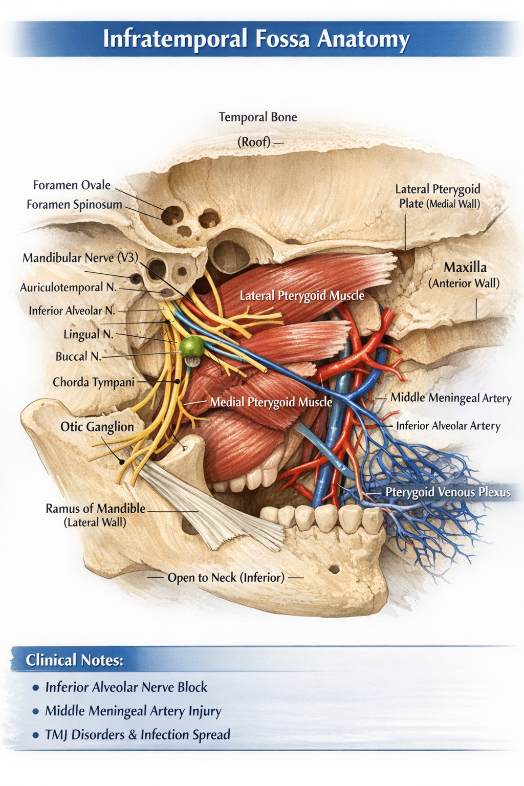
Infratemporal Fossa Anatomy Boundaries Contents Nerves Vessels and Clinical Importance
Frequently Asked Questions
What is the infratemporal fossa?
The infratemporal fossa is an irregular deep anatomical space located below the base of the skull, medial to the ramus of the mandible, and posterior to the maxilla. It contains muscles of mastication, major nerves, vessels, and parasympathetic ganglia.
What forms the roof of the infratemporal fossa?
The roof is formed by the infratemporal surface of the greater wing of the sphenoid and the squamous part of the temporal bone. It contains the foramen ovale and foramen spinosum.
Which nerves pass through the foramen ovale in the infratemporal fossa?
The mandibular nerve (V3), accessory meningeal artery, and the lesser petrosal nerve pass through the foramen ovale.
What are the main contents of the infratemporal fossa?
The main contents include muscles of mastication (medial and lateral pterygoid, lower part of temporalis), mandibular nerve and its branches, maxillary artery and branches, pterygoid venous plexus, chorda tympani, and otic ganglion.
Which artery is most clinically important in the infratemporal fossa?
The middle meningeal artery is clinically important as its injury can cause extradural hematoma.
What is the pterygoid venous plexus and why is it important?
The pterygoid venous plexus is a network of veins in the infratemporal fossa that drains blood from nasal cavity and nasopharynx and communicates with the cavernous sinus, providing a route for spread of infection.
Which muscle is primarily responsible for protrusion of the mandible?
The lateral pterygoid muscle is the primary muscle responsible for protrusion of the mandible.
How does the infratemporal fossa communicate with the pterygopalatine fossa?
The infratemporal fossa communicates with the pterygopalatine fossa through the pterygomaxillary fissure.
What is the function of the otic ganglion?
The otic ganglion provides parasympathetic secretomotor fibers to the parotid gland via the auriculotemporal nerve.
Why is the infratemporal fossa important in dental anesthesia?
The infratemporal fossa is the site where inferior alveolar nerve block is administered for dental procedures, making its anatomy critical for effective and safe anesthesia.
MCQ Test - Infratemporal Fossa Anatomy Boundaries Contents Nerves Vessels and Clinical Importance
Progress:
0/0
Time: 00:00
No MCQs available for this article.