Medical Disclaimer: This is educational content only, not medical advice. Consult a licensed healthcare provider for diagnosis/treatment. Information based on sources like WHO/CDC guidelines (last reviewed: 2026-02-13).
This article is being expanded for more depth. Check back soon!
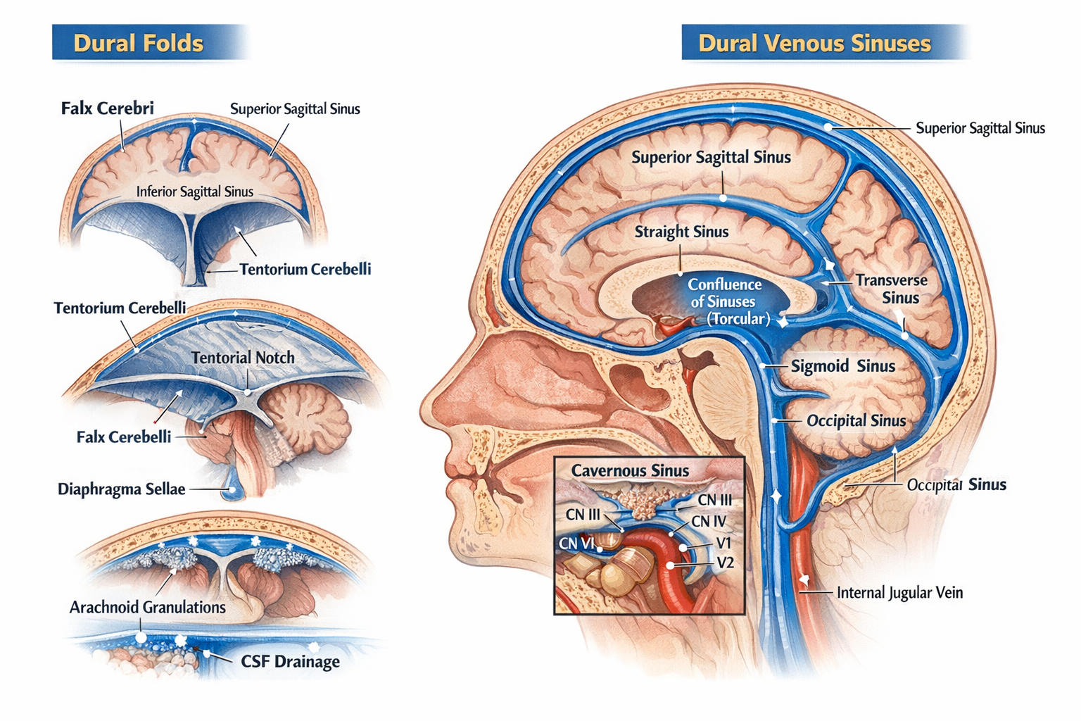
Dural Folds and Dural Venous Sinuses Anatomy, Features and Clinical Significance
Frequently Asked Questions
What are dural folds?
Dural folds are inward reflections of the meningeal layer of dura mater that partition and support the brain within the cranial cavity and help prevent excessive brain movement.
Which are the major dural folds of the brain?
The major dural folds are falx cerebri, tentorium cerebelli, falx cerebelli, and diaphragma sellae.
What is the falx cerebri?
Falx cerebri is a sickle-shaped vertical dural fold that lies in the midline and separates the right and left cerebral hemispheres.
Which dural fold separates the cerebrum from the cerebellum?
The tentorium cerebelli separates the cerebrum above from the cerebellum below.
What is the tentorial notch and why is it important?
The tentorial notch is an opening in the tentorium cerebelli that allows passage of the midbrain; it is clinically important because transtentorial herniation can compress vital brainstem structures.
What is the function of the diaphragma sellae?
The diaphragma sellae forms the roof of the sella turcica and covers the pituitary gland, with a central opening for the pituitary stalk.
What are dural venous sinuses?
Dural venous sinuses are endothelial-lined venous channels located between the periosteal and meningeal layers of dura mater that drain venous blood and cerebrospinal fluid from the brain.
Which sinus lies in the upper margin of the falx cerebri?
The superior sagittal sinus lies in the upper attached margin of the falx cerebri.
Which dural venous sinus receives cerebrospinal fluid?
The superior sagittal sinus receives cerebrospinal fluid through arachnoid villi and arachnoid granulations.
What is the cavernous sinus and why is it clinically significant?
The cavernous sinus is a paired dural venous sinus located on either side of the body of the sphenoid; it is clinically significant because it contains the internal carotid artery and cranial nerves, making it vulnerable to infections and thrombosis.
Which cranial nerve runs within the cavernous sinus?
The abducent nerve (cranial nerve VI) runs within the cavernous sinus alongside the internal carotid artery.
What is the confluence of sinuses?
The confluence of sinuses, also known as torcular Herophili, is the junction where the superior sagittal sinus, straight sinus, and occipital sinus meet.
Which sinus continues as the internal jugular vein?
The sigmoid sinus continues as the internal jugular vein after passing through the jugular foramen.
Why do dural venous sinuses not collapse like normal veins?
Dural venous sinuses do not collapse because they have rigid walls formed by dura mater and lack smooth muscle and valves.
MCQ Test - Dural Folds and Dural Venous Sinuses Anatomy, Features and Clinical Significance
Progress:
0/0
Time: 00:00
No MCQs available for this article.