Medical Disclaimer: This is educational content only, not medical advice. Consult a licensed healthcare provider for diagnosis/treatment. Information based on sources like WHO/CDC guidelines (last reviewed: 2026-02-13).
This article is being expanded for more depth. Check back soon!
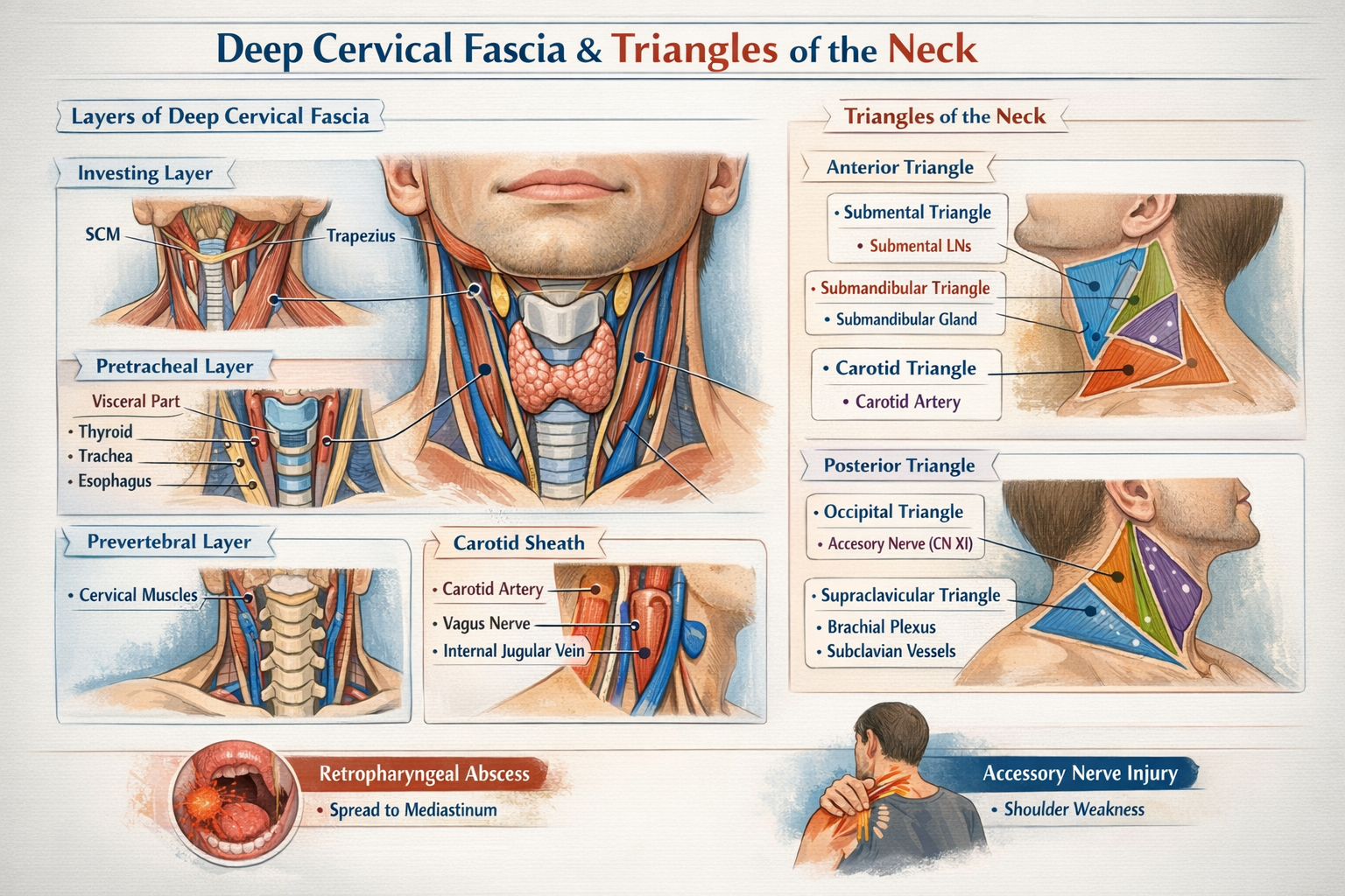
Deep Cervical Fascia and Triangles of the Neck Anatomy Explained in Detail
Frequently Asked Questions
What is the deep cervical fascia?
Deep cervical fascia is a dense connective tissue layer of the neck that encloses muscles, blood vessels, nerves, and viscera, providing support, compartmentalization, and a pathway for the spread or limitation of infections.
What are the main layers of deep cervical fascia?
The main layers are the investing layer, pretracheal layer (muscular and visceral parts), prevertebral layer, and the carotid sheath.
Which muscles are enclosed by the investing layer of deep cervical fascia?
The investing layer encloses the sternocleidomastoid and trapezius muscles.
Which structures are enclosed by the pretracheal fascia?
The pretracheal fascia encloses the infrahyoid muscles (muscular part) and the thyroid gland, trachea, and esophagus (visceral part).
Why does the thyroid gland move during swallowing?
The thyroid gland moves during swallowing because it is attached to the larynx and trachea by the visceral part of the pretracheal fascia, especially through Berry’s ligament.
What structures are contained within the carotid sheath?
The carotid sheath contains the common and internal carotid artery, internal jugular vein, vagus nerve, deep cervical lymph nodes, and sympathetic fibers.
Which fascia forms the axillary sheath?
The prevertebral layer of deep cervical fascia extends laterally to form the axillary sheath enclosing the subclavian artery and brachial plexus.
What is the retropharyngeal space and why is it important?
The retropharyngeal space lies between the buccopharyngeal fascia and alar fascia and is clinically important because infections here can spread to the mediastinum.
How is the neck divided into triangles?
The sternocleidomastoid muscle divides the neck into the anterior triangle and the posterior triangle.
What are the subdivisions of the anterior triangle of the neck?
The anterior triangle is subdivided into the submental, submandibular (digastric), carotid, and muscular triangles.
What are the boundaries of the posterior triangle of the neck?
The posterior triangle is bounded anteriorly by the sternocleidomastoid, posteriorly by the trapezius, inferiorly by the clavicle, and superiorly at the apex where SCM and trapezius meet.
Which nerve is most vulnerable in the posterior triangle of the neck?
The spinal accessory nerve (cranial nerve XI) is most vulnerable as it runs superficially across the posterior triangle.
Which triangle contains the carotid bifurcation and carotid body?
The carotid triangle contains the carotid bifurcation, carotid sinus, and carotid body.
Which triangle provides surgical access to the trachea?
The muscular triangle provides access to the trachea and thyroid gland.
What is the clinical importance of the deep cervical fascia?
Deep cervical fascia directs the spread of neck infections, supports vital structures, forms fascial spaces, and explains clinical features such as painful parotid swelling and mediastinal spread of infections.
MCQ Test - Deep Cervical Fascia and Triangles of the Neck Anatomy Explained in Detail
Progress:
0/0
Time: 00:00
No MCQs available for this article.