This article is being expanded for more depth. Check back soon!

Bundle Branch Block Clinical Features Diagnosis and Management Guide

Author: Medical Editorial Team – Board-certified physicians with 10+ years in emergency medicine. Learn more.

Illustration of Bundle Branch Block Clinical Features Diagnosis and Management Guide symptoms

Medical Disclaimer: This is educational content only, not medical advice. Consult a licensed healthcare provider for diagnosis/treatment. Information based on sources like WHO/CDC guidelines (last reviewed: 2026-02-13).

About the Author: Dr. Dinesh, MBBS, is a qualified medical doctor with over [2 years – add your experience] of experience in general medicine As the owner and lead content creator of LearnWithTest.pro, Dr. Dinesh ensures all articles are based on evidence-based guidelines from sources like WHO, CDC, and peer-reviewed journals. This content is for educational purposes only and not a substitute for professional medical advice.

Contact Details: For inquiries or collaborations, reach out at:

All content is reviewed for accuracy and updated regularly (last review: January 10, 2026). We prioritize trustworthiness by citing reliable sources and adhering to medical ethics.

Frequently Asked Questions

What is a bundle branch block?

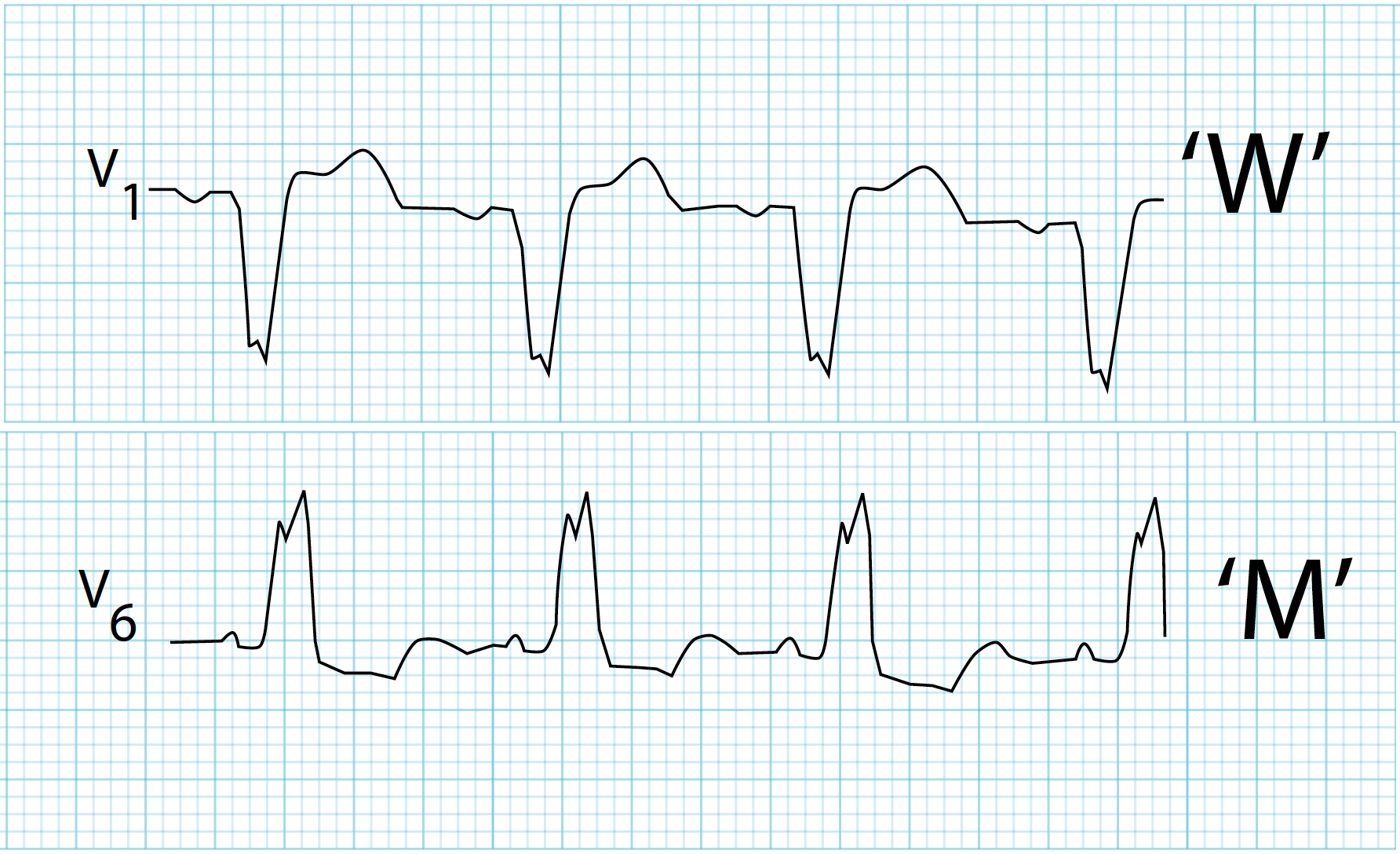
A bundle branch block is a cardiac conduction abnormality in which electrical impulses are delayed or blocked in the right or left bundle branch of the His–Purkinje system, resulting in widened QRS complexes and asynchronous ventricular depolarization on ECG.

What is the difference between right and left bundle branch block?

Right bundle branch block affects right ventricular depolarization and is often benign, while left bundle branch block affects left ventricular depolarization and is usually associated with underlying structural heart disease and worse prognosis.

What are the common causes of left bundle branch block?

Common causes include hypertension with left ventricular hypertrophy, ischemic heart disease, cardiomyopathy, aortic stenosis, myocardial infarction, and degenerative conduction system disease.

Is bundle branch block always symptomatic?

No, many patients with bundle branch block are asymptomatic. Symptoms such as syncope, dizziness, or dyspnea usually indicate associated heart disease or progression to advanced conduction block.

Why is new-onset left bundle branch block with chest pain considered dangerous?

New-onset left bundle branch block can mask ischemic ECG changes and is treated as a STEMI equivalent because it often represents acute myocardial infarction requiring urgent reperfusion.

How is bundle branch block diagnosed?

Diagnosis is made using a 12-lead ECG showing QRS duration of 120 ms or more with characteristic morphologic patterns specific to right or left bundle branch block.

Does right bundle branch block require treatment?

Isolated asymptomatic right bundle branch block usually does not require treatment. Management focuses on identifying and treating any underlying cardiac or pulmonary condition.

When is a pacemaker indicated in bundle branch block?

A pacemaker is indicated in patients with bundle branch block who develop symptomatic high-grade atrioventricular block, recurrent syncope due to conduction disease, alternating bundle branch block, or trifascicular disease.

What is the role of cardiac resynchronization therapy in bundle branch block?

Cardiac resynchronization therapy is recommended in patients with heart failure, reduced ejection fraction (≤35%), left bundle branch block morphology, and wide QRS duration to improve symptoms and survival.

What is the prognosis of bundle branch block?

Prognosis depends on the type and underlying cause. Isolated right bundle branch block has a good prognosis, while left bundle branch block is associated with increased risk of heart failure, arrhythmias, and cardiovascular mortality.

MCQ Test - Bundle Branch Block Clinical Features Diagnosis and Management Guide

Progress:
0/15
Time: 00:00

1 A 64-year-old man with known ischemic cardiomyopathy presents with worsening dyspnea, orthopnea, and bilateral basal crepitations. Echocardiography shows LVEF 28%. Which drug provides the greatest mortality benefit in this patient?

Explanation:

ACE inhibitors reduce mortality and morbidity in heart failure with reduced ejection fraction by inhibiting maladaptive RAAS activation and preventing ventricular remodeling.

2 A 70-year-old hypertensive woman presents with exertional dyspnea. Echocardiography shows concentric LV hypertrophy with preserved ejection fraction of 60%. What is the most likely diagnosis?

Explanation:

Preserved ejection fraction with diastolic dysfunction and concentric hypertrophy is characteristic of HFpEF.

3 A patient with chronic heart failure develops sudden weight gain of 3 kg over 4 days, ankle edema, and raised JVP. What is the primary mechanism responsible?

Explanation:

RAAS activation leads to sodium and water retention, resulting in congestion and rapid weight gain.

4 A patient with stable HFrEF is started on a beta blocker and develops transient worsening of dyspnea in the first week. What is the most likely explanation?

Explanation:

Beta blockers initially reduce myocardial contractility, which can transiently worsen symptoms before long-term benefit occurs.

5 A patient with acute decompensated heart failure presents with cold extremities, hypotension, oliguria, and pulmonary edema. Which hemodynamic profile best describes this patient?

Explanation:

Cold and wet profile indicates low cardiac output with volume overload and is associated with poor prognosis.

6 A patient with CHF has serum potassium of 6.0 mmol/L and rising creatinine. Which drug should be stopped first?

Explanation:

Mineralocorticoid receptor antagonists commonly cause hyperkalemia and should be withheld first.

7 A patient with CHF and atrial fibrillation remains symptomatic despite optimal therapy. Which drug is most useful for symptom control and ventricular rate control?

Explanation:

Digoxin improves symptoms and provides rate control in atrial fibrillation with heart failure.

8 A patient with heart failure has raised JVP, hepatomegaly, ascites, and pedal edema. Which ventricle is primarily failing?

Explanation:

Systemic venous congestion indicates predominant right-sided heart failure.

9 A patient with suspected heart failure has normal BNP levels. What is the most appropriate interpretation?

Explanation:

Normal BNP or NT-proBNP levels make heart failure unlikely in most clinical settings.

10 A patient with HFrEF is switched from enalapril to sacubitril–valsartan. What precaution is mandatory?

Explanation:

A 36-hour washout after stopping ACE inhibitors is required to prevent angioedema.

11 A patient with CHF has an S3 gallop on auscultation. What does this finding indicate?

Explanation:

S3 gallop is associated with increased ventricular filling pressures and volume overload.

12 A patient with chronic heart failure and LVEF 30% remains symptomatic despite optimal therapy. Which device reduces sudden cardiac death risk?

Explanation:

ICD therapy reduces the risk of sudden cardiac death in patients with LVEF 35% or less.

13 A patient with CHF develops worsening renal function after aggressive diuretic therapy. What is this condition called?

Explanation:

Cardiorenal syndrome reflects the bidirectional interaction between heart failure and renal dysfunction.

14 A patient with HFpEF presents with exertional dyspnea. Which management strategy is most important?

Explanation:

Management of HFpEF focuses on blood pressure control and treatment of comorbid conditions.

15 A patient with end-stage congestive heart failure remains symptomatic despite maximal medical therapy. What is the most definitive treatment option?

Explanation:

Advanced heart failure requires mechanical circulatory support or heart transplantation for definitive management.

Test Results

0%
0/15
0
Correct Answers
0
Wrong Answers
00:00
Time Taken
0
Skipped
View Exam Mode MCQs (1767531084-index-2026-01-04T182031.468.html)

Related Articles

Comments