This article is being expanded for more depth. Check back soon!

Pulseless Electrical Activity Clinical Features Diagnosis and Management Guide

Author: Medical Editorial Team – Board-certified physicians with 10+ years in emergency medicine. Learn more.

Illustration of Pulseless Electrical Activity Clinical Features Diagnosis and Management Guide symptoms

Medical Disclaimer: This is educational content only, not medical advice. Consult a licensed healthcare provider for diagnosis/treatment. Information based on sources like WHO/CDC guidelines (last reviewed: 2026-02-13).

About the Author: Dr. Dinesh, MBBS, is a qualified medical doctor with over [2 years – add your experience] of experience in general medicine As the owner and lead content creator of LearnWithTest.pro, Dr. Dinesh ensures all articles are based on evidence-based guidelines from sources like WHO, CDC, and peer-reviewed journals. This content is for educational purposes only and not a substitute for professional medical advice.

Contact Details: For inquiries or collaborations, reach out at:

All content is reviewed for accuracy and updated regularly (last review: January 10, 2026). We prioritize trustworthiness by citing reliable sources and adhering to medical ethics.

Frequently Asked Questions

What is pulseless electrical activity (PEA)?

Pulseless electrical activity is a cardiac arrest rhythm in which organized electrical activity is seen on ECG but there is no palpable pulse or effective cardiac output.

Is pulseless electrical activity a shockable rhythm?

No, pulseless electrical activity is a non-shockable rhythm and should not be treated with defibrillation.

What is the main cause of death in pulseless electrical activity?

Death in PEA occurs due to failure of mechanical cardiac contraction despite preserved electrical activity, leading to complete circulatory collapse.

What are the most common reversible causes of PEA?

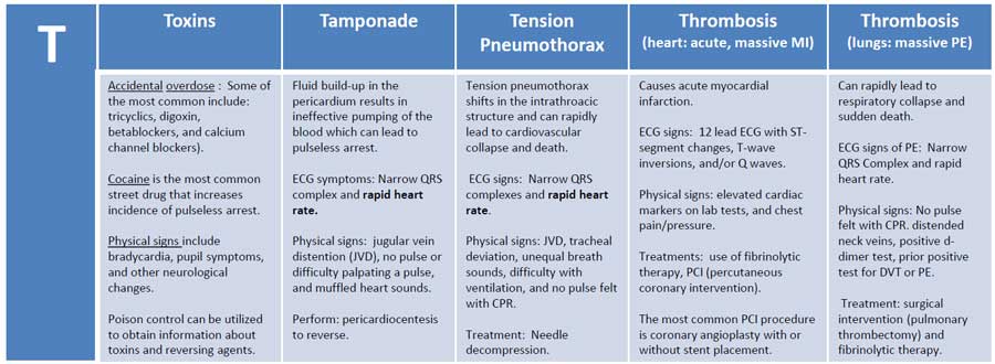
The common reversible causes are summarized as Hs and Ts, including hypovolemia, hypoxia, acidosis, hypo- or hyperkalemia, hypothermia, tension pneumothorax, cardiac tamponade, toxins, thrombosis (coronary or pulmonary), and trauma.

What is the first-line treatment for pulseless electrical activity?

Immediate high-quality CPR along with early administration of intravenous or intraosseous epinephrine and rapid identification of reversible causes.

What is the recommended dose of epinephrine in PEA?

The recommended adult dose is 1 mg IV or IO every 3–5 minutes during resuscitation.

Why is defibrillation not used in PEA?

Defibrillation is ineffective because PEA does not involve chaotic electrical activity but rather a failure of mechanical contraction.

What is pseudo-PEA?

Pseudo-PEA refers to organized electrical activity with minimal cardiac contraction detectable only by ultrasound, without a palpable pulse.

How does ultrasound help in managing PEA?

Bedside ultrasound helps identify reversible causes such as cardiac tamponade, massive pulmonary embolism, severe hypovolemia, and distinguishes true PEA from pseudo-PEA.

Which laboratory abnormality commonly causes PEA in renal failure patients?

Severe hyperkalemia is a frequent and life-threatening cause of PEA in patients with renal failure.

What role does end-tidal CO2 play during PEA resuscitation?

End-tidal CO2 monitoring helps assess CPR quality and prognosis, with persistently low values indicating poor perfusion and low chance of ROSC.

What is the prognosis of pulseless electrical activity compared to shockable rhythms?

PEA generally has a poorer prognosis than ventricular fibrillation or pulseless ventricular tachycardia, especially if no reversible cause is identified.

Can myocardial infarction present as PEA?

Yes, massive myocardial infarction or mechanical complications such as ventricular rupture can lead to PEA.

What is the most important principle in treating PEA?

The most important principle is rapid identification and correction of reversible causes while continuing high-quality CPR.

When should resuscitation be terminated in PEA?

Termination is considered when prolonged high-quality resuscitation fails to achieve ROSC and no reversible causes are identified, following institutional and ethical guidelines.

MCQ Test - Pulseless Electrical Activity Clinical Features Diagnosis and Management Guide

Progress:
0/15
Time: 00:00

1 A 62-year-old man collapses in the emergency department. ECG shows organized sinus rhythm at 80/min, but no carotid pulse is palpable. What is the most appropriate immediate action?

Explanation:

Organized electrical activity without a pulse defines PEA, which requires immediate CPR and epinephrine, not defibrillation.

2 A trauma patient develops PEA arrest. Neck veins are distended and breath sounds are absent on the right side. What is the most likely cause?

Explanation:

Distended neck veins with unilateral absent breath sounds strongly suggest tension pneumothorax causing PEA.

3 A post–myocardial infarction patient develops sudden PEA. Bedside ultrasound shows a large pericardial effusion with diastolic chamber collapse. What is the definitive management?

Explanation:

Cardiac tamponade is a reversible cause of PEA and requires urgent pericardiocentesis.

4 A chronic kidney disease patient misses dialysis and presents with PEA arrest. Which ECG finding before arrest most likely preceded this rhythm?

Explanation:

Hyperkalemia, common in renal failure, produces peaked T waves and can precipitate PEA.

5 During CPR for PEA, end-tidal CO₂ remains persistently below 5 mmHg. What does this indicate?

Explanation:

Persistently low ETCO₂ reflects inadequate perfusion and is associated with poor outcomes.

6 A patient with sudden onset dyspnea collapses with PEA. Ultrasound shows dilated right ventricle and septal bowing. What is the most likely diagnosis?

Explanation:

Acute right ventricular dilation with septal shift in PEA strongly suggests massive pulmonary embolism.

7 A patient in septic shock progresses to PEA arrest. What is the primary pathophysiologic mechanism?

Explanation:

Sepsis causes profound vasodilation and myocardial dysfunction, leading to PEA.

8 A patient with beta-blocker overdose develops PEA. Which drug is most appropriate as targeted therapy?

Explanation:

Glucagon bypasses beta-adrenergic receptors and improves myocardial contractility in beta-blocker toxicity.

9 A pregnant woman in late third trimester develops PEA arrest. Which immediate maneuver improves venous return?

Explanation:

Left uterine displacement relieves aortocaval compression and improves preload during resuscitation.

10 Despite repeated epinephrine doses, PEA persists. What is the most important next step?

Explanation:

Correction of reversible causes (Hs and Ts) is the cornerstone of successful PEA management.

11 Ultrasound during PEA shows minimal cardiac activity without a palpable pulse. What is this condition called?

Explanation:

Pseudo-PEA has minimal cardiac contraction detectable only on ultrasound.

12 A trauma patient with massive blood loss develops PEA. Which intervention is most critical?

Explanation:

Hypovolemia is a leading cause of traumatic PEA and requires rapid volume replacement.

13 What is the primary benefit of epinephrine in PEA resuscitation?

Explanation:

Alpha-adrenergic vasoconstriction increases coronary and cerebral perfusion pressure.

14 After ROSC following PEA arrest, which measure is most important?

Explanation:

Post-ROSC care focuses on maintaining perfusion and treating the underlying cause.

15 Persistent true PEA with cardiac standstill on ultrasound despite prolonged resuscitation suggests what prognosis?

Explanation:

Persistent cardiac standstill in true PEA is associated with very low survival rates.

Test Results

0%
0/15
0
Correct Answers
0
Wrong Answers
00:00
Time Taken
0
Skipped
View Exam Mode MCQs (1767616574-index-2026-01-05T180511.960.html)

Related Articles

Comments