Medical Disclaimer: This is educational content only, not medical advice. Consult a licensed healthcare provider for diagnosis/treatment. Information based on sources like WHO/CDC guidelines (last reviewed: 2026-02-13).
Aortic Stenosis Complete Clinical Guide Causes Symptoms Diagnosis Treatment
Frequently Asked Questions
What is aortic stenosis?
Aortic stenosis is a valvular heart disease characterized by narrowing of the aortic valve opening, leading to obstruction of left ventricular outflow, increased pressure load on the left ventricle, and reduced cardiac output.
What are the most common causes of aortic stenosis?
The most common causes are degenerative calcific aortic stenosis in elderly patients, bicuspid aortic valve in younger individuals, and rheumatic heart disease in endemic regions.
What are the classic symptoms of severe aortic stenosis?
The classic triad includes exertional angina, syncope or presyncope, and dyspnea due to heart failure. Symptoms usually indicate advanced disease and poor prognosis without intervention.
Why does angina occur in aortic stenosis even without coronary artery disease?
Angina occurs due to increased myocardial oxygen demand from left ventricular hypertrophy and reduced coronary perfusion reserve caused by elevated intraventricular pressures.
What are the key physical examination findings in aortic stenosis?
Findings include a harsh ejection systolic murmur at the right upper sternal border radiating to the carotids, pulsus parvus et tardus, narrow pulse pressure, soft or absent A2, and a sustained heaving apex beat.
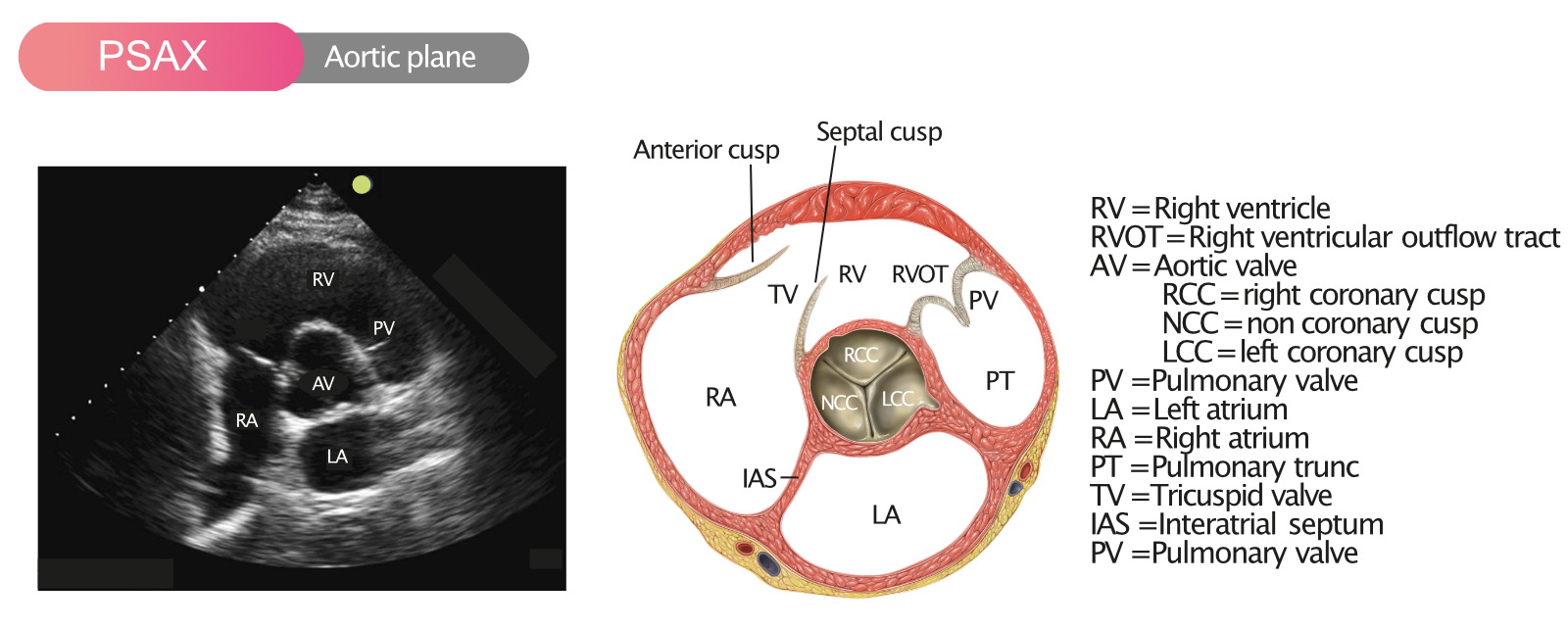
How is the severity of aortic stenosis assessed on echocardiography?
Severity is assessed using peak aortic jet velocity, mean transvalvular gradient, and aortic valve area calculated by the continuity equation.
What defines severe aortic stenosis on echocardiography?
Severe aortic stenosis is defined by aortic valve area ≤ 1.0 cm², peak velocity ≥ 4.0 m/s, or mean gradient ≥ 40 mmHg.
What is low-flow, low-gradient aortic stenosis?
It is a subtype of severe aortic stenosis where transvalvular gradients are low due to reduced stroke volume, often seen with reduced ejection fraction or paradoxically with preserved ejection fraction.
Which investigation helps differentiate true severe from pseudo-severe aortic stenosis?
Low-dose dobutamine stress echocardiography is used to assess contractile reserve and changes in valve area to distinguish true severe from pseudo-severe aortic stenosis.
Is medical therapy sufficient for severe aortic stenosis?
No, medical therapy does not halt disease progression or improve survival in severe aortic stenosis. Definitive treatment requires aortic valve replacement.
When is aortic valve replacement indicated in aortic stenosis?
It is indicated in all patients with severe aortic stenosis who develop symptoms or left ventricular systolic dysfunction, and in selected high-risk asymptomatic patients.
What are the main types of aortic valve replacement?
The two main types are surgical aortic valve replacement (SAVR) and transcatheter aortic valve replacement (TAVR).
Which patients are preferred candidates for TAVR?
Elderly patients, those with high or prohibitive surgical risk, and selected intermediate-risk patients after heart team evaluation are preferred candidates for TAVR.
What is the role of balloon aortic valvuloplasty?
Balloon aortic valvuloplasty is used as a temporary bridge to definitive valve replacement or as palliative therapy in patients who are not candidates for SAVR or TAVR.
What complications can occur if aortic stenosis is left untreated?
Complications include heart failure, atrial fibrillation, ventricular arrhythmias, sudden cardiac death, pulmonary hypertension, and gastrointestinal bleeding due to Heyde syndrome.
What is Heyde syndrome?
Heyde syndrome is the association of severe aortic stenosis with gastrointestinal angiodysplasia and acquired von Willebrand factor deficiency, leading to recurrent GI bleeding.
Why is atrial fibrillation poorly tolerated in aortic stenosis?
Patients with aortic stenosis rely heavily on atrial contraction for left ventricular filling, and loss of atrial kick can rapidly precipitate heart failure.
What is the prognosis of symptomatic severe aortic stenosis without valve replacement?
The prognosis is poor, with a median survival of approximately 2 to 3 years once symptoms develop.
How should asymptomatic patients with severe aortic stenosis be followed?
They require close clinical monitoring, periodic echocardiography, and exercise testing in selected cases to detect early symptom development or disease progression.
Can aortic stenosis be prevented?
There is no proven therapy to prevent degenerative aortic stenosis, but controlling cardiovascular risk factors and early detection in bicuspid valve disease may help delay complications.
MCQ Test - Aortic Stenosis Complete Clinical Guide Causes Symptoms Diagnosis Treatment
Progress:
0/15
Time: 00:00
Test Results
0%
0/15
0
Correct Answers
0
Wrong Answers
00:00
Time Taken
0
Skipped