Medical Disclaimer: This is educational content only, not medical advice. Consult a licensed healthcare provider for diagnosis/treatment. Information based on sources like WHO/CDC guidelines (last reviewed: 2026-02-13).
First Heart Sound S1 and Jugular Venous Pressure JVP Complete Clinical Guide
Frequently Asked Questions
What is the first heart sound S1?
The first heart sound S1 is produced mainly by closure of the mitral and tricuspid valves at the onset of ventricular systole and coincides with the carotid pulse and R wave of ECG.
What are the components of S1?
S1 consists of two components: M1 from mitral valve closure and T1 from tricuspid valve closure, which are usually heard as a single sound.
What causes a loud S1?
A loud S1 occurs when AV valve leaflets are wide open at the start of systole, commonly seen in mitral stenosis with mobile leaflets, short PR interval, tachycardia, and hyperdynamic states.
What causes a soft S1?
A soft S1 is seen in mitral regurgitation, long PR interval, left ventricular dysfunction, and calcified or immobile mitral valves.
Why does S1 vary in intensity in atrial fibrillation?
In atrial fibrillation, variable diastolic filling leads to inconsistent ventricular contraction force, resulting in beat-to-beat variation in S1 intensity.
What is jugular venous pressure JVP?
Jugular venous pressure is a clinical estimate of right atrial pressure assessed by observing pulsations of the internal jugular vein.
Why is the internal jugular vein used to assess JVP?
The internal jugular vein has a direct connection to the right atrium without valves, making it a reliable indicator of right atrial pressure.
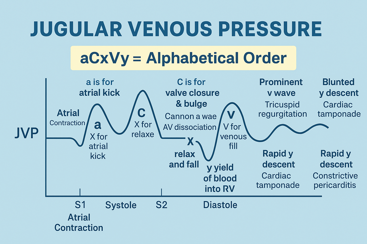
What are the normal components of the JVP waveform?
The normal JVP waveform includes the a wave, c wave, x descent, v wave, and y descent, reflecting different phases of the cardiac cycle.
What causes absent a waves in JVP?
Absent a waves are seen in atrial fibrillation due to loss of organized atrial contraction.
What are cannon a waves and when are they seen?
Cannon a waves are large intermittent a waves caused by atrial contraction against a closed tricuspid valve, seen in complete heart block and other forms of AV dissociation.
What is Kussmaul sign?
Kussmaul sign is a paradoxical rise in JVP during inspiration, seen in constrictive pericarditis, restrictive cardiomyopathy, and right ventricular infarction.
What is the clinical significance of raised JVP?
Raised JVP indicates elevated right atrial pressure and is commonly seen in right heart failure, tricuspid valve disease, pericardial disease, and fluid overload.
MCQ Test - First Heart Sound S1 and Jugular Venous Pressure JVP Complete Clinical Guide
Progress:
0/15
Time: 00:00
Test Results
0%
0/15
0
Correct Answers
0
Wrong Answers
00:00
Time Taken
0
Skipped